|
|||||
| If you just print this out, you'll miss some of its function. As you review it online, you can put the cursor on parts of the heart and they will be identified for you. | |||||
|
|||||
Heart Dissection instructions - external |
|||||
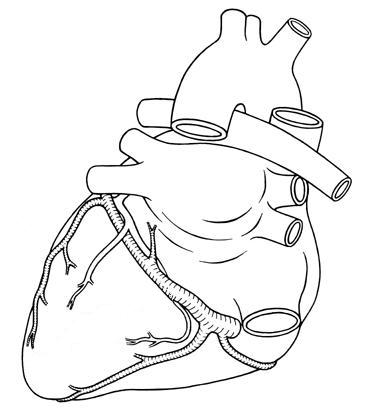
Remove the outer parietal pericardium (if present) by cutting it from the apex of the heart to the base with scissors and gently peeling it off.
The right ventricle and right atrium are separated by a groove, the coronary sulcus. Find the right coronary artery running down this groove. Find the auricles, the hollow ‘earlike’ extension of the atria. On the posterior side of the heart you can see the left atrium. Pulmonary veins usually are cut off during removal of the heart and are often only visible as holes in the left atrium. The coronary sulcus continues around the posterior side of the heart, where it separates the left atrium from the left venticle. You can see the great coronary vein running along this sulcus. |
|||||
 |
|||||

 |
|
Heart dissection instructions - internal |
|
| Cut down the vena cava into the right atrium to expose the right AV valve (tricuspid valve).. Fill the right ventricle with water, pouring it in through the right AV valve. Gently squeeze the walls of the ventricle to note the closing action of the valve’s cusps. Look at the atrial septum, the wall between left and right atria, and see if you can find a roundish scar where the foramen ovale closed. This was an opening between the atria during fetal life. Drain the water from the right ventricle and continue the cut with scissors from the right atrium through the right AV valve down to the apex of the heart. Expose the right ventricle and locate the papillary muscles and chordae tendineae in the right ventricle. Cut upwards along the ventricular and atrial septa towards the base of the heart to expose the pulmonary semilunar valve. Which direction does blood flow through this valve? |
|

|
||||
Insert one blade of your scissors into the left atrium. Cut through the left atrium into the left ventricle to the apex of the heart. Note the two leaflets of the left AV valve (bicuspid or mitral valve). Also cut upwards from the left ventricle into the aortic arch to expose the aortic semilunar valve and the two openings to the coronary arteries. Modified from Andrews, 2007. http://www.biol.andrews.edu/anat/anp2/lab/anp2.l2heartb.html |
||||
| Use these vocabulary roots to answer the questions below: | ||||
| brady- = slow tachy- = fast a- = without anti= - against cardio- = referring to the heart angio- = referring to the blood vessels and lymph vessels -megaly = enlargement athero- or arterio- = referring to the arteries veno- or phleb- = refering to the veins sclerosis = hardening of a tissue, usually by fibrous deposits with calcium in them |
||||
| bradycardia = ___________________________________ A fast heartbeat would be __________________________________ Asystole = _________________________________________ Phlebitis = _______________________________ An antiarrhythmic drug would be used to: __________________________________________________ Angiopathy would be ___________________________________ The study of blood vessels would be ______________________________ a tumor composed of blood vessels would be an _______________________________ Atherosclerosis = ___________________________ Acardius = _________________________________ Venopuncture = ______________________________ |
||||
| Quantitative Literacy! | ||||
You know systole and diastole. Now use these concepts: Cardiac Output (CO) = heart rate x stroke volume SHOULD BE 3-6 L/min
Pulse pressure = the difference between systolic and diastolic pressures |
||||
A man has been brought into the ICU after cardiac surgery. Fill in the chart:: HR = 94 bpm Weight 204 lb CO = ________________________________ CI = _________________________________ PP = _________________________________ MAP = _______________________________ SVR = ________________________________ Is this patient doing OK, or would you call his doctor? Give your rationale. |
 |
|||